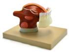
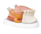
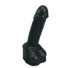
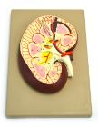
Enlarged 3x, shows the male urinary bladder with the prostate gland surrounding the urethra. The model is dissected medially to expose both inteRNAl and exteRNAl structures of the bladder and prostate
Model Human Urinary Organs, Weight (lb):2.6, Length (in):13.0,Width (in): 9.1, Height (in): 4.3,Natural Size separates into 3 parts. Kidneys ureters, adrenal glands, bladder with prostate and major blood vessels
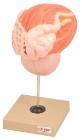
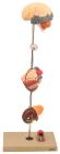
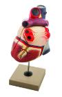
Showing the harmful effects of hypertension on the most susceptible organs. It consists of scaled down depictions of brain, eye, 2-part heart, 2-part kidney, enlarged artery. All of the organs can be rotated or removed for closer viewing.
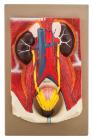
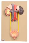
Natural size. Kidneys, ureters, adrenal glands and bladder with prostate as well as the large abdominal right kidney sectioned to show all anatomical details.
Life size model dissectible in 2 parts. The anterior heart wall can be removed to show the left and right ventricles and atria as well as the tricuspid, pulmonary, mitral and aortic valves. Mounted on base.
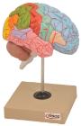
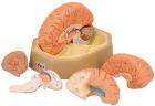
Life size, half of human brain. Students can study regions within the cerebral cortex are allied to certain functions. Impulses from the sensory organs, the skeletal muscles, skin and joints all travel to areas specialized in interpreting the information
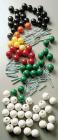
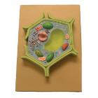
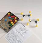
This plant cell model is separated in 4 parts. Showing details of cell wall and inner details of cell wall, nucleus is separated and defined in a 3-D way cut section of chloroplast is shown.
Amoeba proteus, enlarged approximately 1000 times. In a small pseudopodium which can be opened up showing the structure after electron microscopic magnification. On a base with explanatory notes. Separates into 2 parts
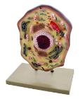
Enlarged 20,000 times. The model shows delicate structure of an animal cell. Features organelles nucleus, endoplasmic reticulum, mitochondria, ribosomes respectively polysomic and Golgi apparatus. Also showing centrioles, lysosomes and fat vacuoles.
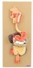
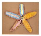
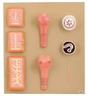
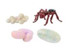
Ant Life Cycle Stages, Children can see how ants change as they grow with Insect Lore’s Ant Life Cycle Stages. These oversized, anatomically correct figures have been accurately painted and sculpted to show the four stages of ant development: eggs, larva, pupa and adult.
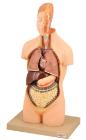
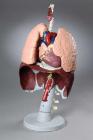
Model Respiratory Organ, unbreakable life-size model is distinctively colored to illustrate anatomical detail. Includes diaphragm, aorta origins of five abdominal arteries, three thoracic and lumbar vertebrae, sectioned left lung, Dimension: 16x9x71/2 in
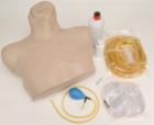
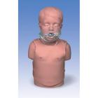
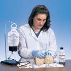
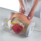
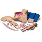
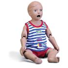
Birthing Simulator Basic, for the skill training in normal deliveries, in complicated deliveries and in obstetric emergencies, Obstetric simulation has proven successful to enhance the training of delivery skills, following of protocols and reaction in emergency situation
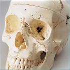
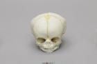
Model Fetal Human Skull 35 Weeks, Scientific Name: Homo sapiens, skull exhibits characteristics of prenatal development, 1-part skull (jaw glued to cranium), Size: 9.7L x 8.2W x 7.5H (cm)
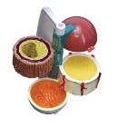
Longitudinal section of the right kidney. Model shows kidney glomerulus, tubes, one collection tube, pyramids, kidneys orifice system, kidney pelvis, upper section of the ureter and the kidney blood vessels
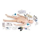
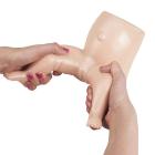
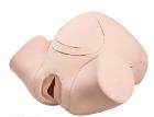
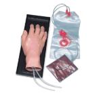
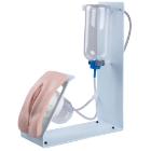
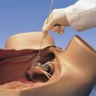
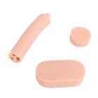
Catheterization Simulator Basic, With the female P93, Female catheterization with realistic resistance, Soft and movable labia, Liquid outflow if catheterization is successfully carried out, The situation can be checked through the transparent bladder
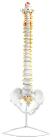
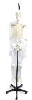
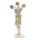
Mini-Skeleton with Flexible Spine, flexible spine to demonstrate natural movement of spine and thorax. Ideal for the chiropractor and orthopedic surgeon. Bones of the skeleton are individually represented - results in full articulation and movements of joints
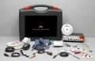
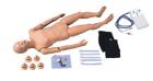
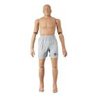
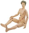
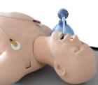
ADAM-X is the male patient simulator designed for practicing skills and providing medical assistance in case of emergency, reproduction of a skeletal and anatomical structure of a human
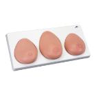
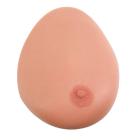
Model Single Breast, With Benign Tumor, Dimensions: 25.4 x 17.8 x 17.8 cm, made of 3B SKINlike* silicone with simulated benign tumour for the demonstration of ultrasonic B-image mode with Ultrasonic Echoscope GS200