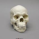
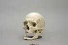
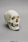
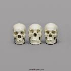
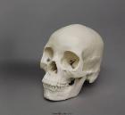
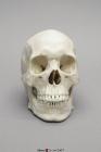
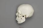
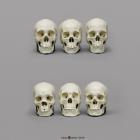
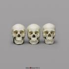
Model Human Male African-American Skull, a depressed nasal root, obtuse nasal angle, short anterior nasal spine, bilateral gutter at lower part of nasal aperture, rectangular-shaped palate & blade-like incisor in upper jaw, Size: 20.9x13.7x20cm
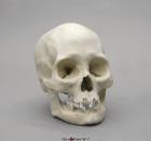
Model Human Elderly European Male Skull (80 years old), Features of advanced age are limited to marked extra & intracranial suture closure, irregular bony exostosis & further suggested by edentulous nature of jaws, Size: 20.4x13.7x17.6cm
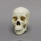
Model Human Female Asian Skull, include moderate alveolar prognathism, flat nasal region, round/square anterior nasal aperture, lack of a nasal sill, no nasal gutter, round maxillary arcade, and short nasal spine, Size: 20.5L x 12.3W X 18.7H (cm)
Model Human Female Asian Skull, include a flat nasal root, an obtuse nasal angle, a somewhat rounded dental arcade in the upper jaw, mild to moderate prognathism and shovel-like incisors in the upper jaw, Size: 19.4L x 12.9W X 17.3H (cm)
Model Human Female American Indian Skull, Includes broad, flattened, forward-projecting zygomas, facial bones are vertically aligned with shallow nasal depression, moderate nasal spine, orthognathic jaw & vertical chin, Size: 18.5x14x18.8cm
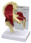
Human Male Polynesian Skull, sagittal keel, parietal bossing, the broad, prominent basiooccipital, and the rocker jaw, rocker jaw is curved along the inferior surface of the mandible, 2-part skull (separate cranium & jaw), Size: 20.7x14.2x19.6cm
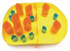
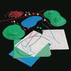
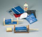
Mitosis & Meiosis 3-D Model Kit-10 Student Model Template Sets and Teacher Guide, Vinyl Pouch, Assemble and use 3-D models to visualize and investigate key science structures, Perfect for use in the classroom, lab or at home, Satisfies NGSS Standards, Grade: 6-10, Material: Paper, LxW: 12X19in
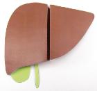
Liver and Spleen 3-D Model Kit-10 Student Model Template Sets and Teacher Guide, Vinyl Pouch, Assemble and use 3-D models to visualize and investigate key science structures, Perfect for use in the classroom, lab or at home, Satisfies NGSS Standards, Grade: 6-10, Material: Paper, LxW: 12X19in
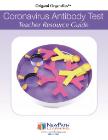
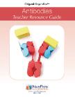
Antibodies 3-D Model Kit-10 Student Model Template Sets and Teacher Guide, Vinyl Pouch, Assemble and use 3-D models to visualize and investigate key science structures, Perfect for use in the classroom, lab or at home, Satisfies NGSS Standards, Grade: 6 to 10, Material: Paper, LxW: 12X19in
Model Human Female Asian Economy Skull, Features no cranial sites for musculofascial attachment, a narrow ascending mandibular ramus, smooth nasion, sharp supraorbital margins & a rounded inferior border of mandible, Size: 17.1x12x16.5cm
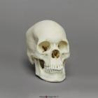
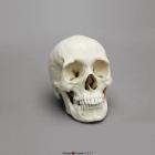
Model Human Female European Skull, generally gracile, specimen is not definitively female, serve as a good discussion piece in a classroom setting for the diagnostic limitations in the determination of sex, Size: 19.2L x 12.6W x 19H (cm)
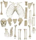
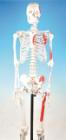
Half Skeleton, unmounted, like Disarticulated Skeleton, but only with ribs and the bones of upper and lower limb of one side of the skeleton. Hand and foot mounted on nylon or wire or unmounted, Dimension and weight: 92x38x26cm, 4.5kg
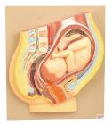
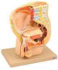
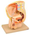
Life size, 19 parts, half of brain, eye with muscles and optic nerve, 2-lung halves, heart in 2 parts, liver, stomach, small/large intestines, kidney, bladder with female pelvic floor and first lumbar vertebra.
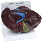
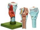
Life size model showing nose, mouth cavity and pharynx, liver with gall bladder, pancreas and spleen. Transverse colon and front stomach wall are removable. Mounted on base. Numbered with Key Card
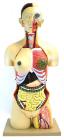
Torso model, hand painted, consists of 13 parts, removeable parts: eye with optical nerve, half brain, lung (2 parts), heart (2 parts), liver, stomach, front portion of the kidney, half of the bladder, large and small intenstines, with appendix flap opening, All parts labeled
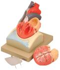
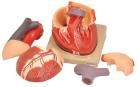
Enlarged, sectioned so that both ventricles and atria open to expose the valves. Large blood vessels near the heart and musculature of the heart are shown. Separates into 4 parts. On base.
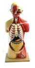
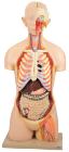
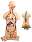
Life size torso model with 28 parts for detailed study of all major organ systems. Dual sex with an open back.Rib cage removed to expose removable inteRNAl organs for detailed study.
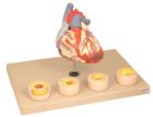
Heart dissected in two parts shows the thickened ventricle walls and cardiac valves. Four individual blood vessel model show coronary artery disease/ atherosclerosis and progression in the blood vessel, myocardial infarction and damage.