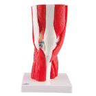
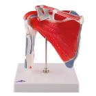
Model Knee Joint 12-Part, with removable joints is part of a high quality series of models, manufactured to precise anatomical correctness with realistic colors of the joint, bone and muscles, extremely durable, nonhazardous material of standards, Dimensions : 33x17x17 cm
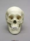
Model Human Adolescent Skull, features of race are of Asian, the totality of features is most with those of slightly gracile young male who has not yet fully developed his sexual characteristics & might be of a slightly robust female, Size: 19x14.2x20.1cm
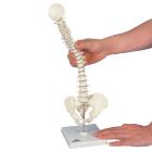
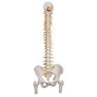
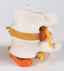
Model Mini Vertebral Column With Stand, with squama occipitalis and pelvis, mounted flexibly to demonstrate natural movements and pathological changes, Weight: 0.6 kg, Dimensions : 44 cm
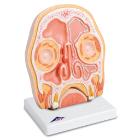
Model Head Section, Frontal section through the human paranasal sinuses covered with mucous membrane. Signs of sinusitis (paranasal sinus inflammation) on the right, with normal ventilation of the left side, Weight: 1.45 kg, Dimensions : 41 x 31 x 5 cm
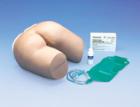
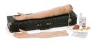
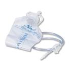
Sim Cathi Catheterization Model, Cathi is a simulated female catheterization trainer, Cathi fits into most Laerdal manikins or into the Remedy Stand, Tissue is supple and pliable for easy spreading using the thumb and index finger, Bladder module is used with IV bag
Ribs, half set, 12 pcs, Model, Human Bones, Plastic, These individual bones are a great addition to you collection, used as a match for your existing Eisco disarticulated skeleton, or to allow students to compare general skeletal structure
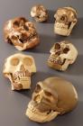
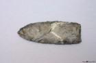
Model Farpoint Clovis Point, 11,000 YA. 5-1/4-in Farpoint Clovis spearpoint was found by Edgar Perez in 2005, Malibu, first such artifact found on the West Coast, indicates the presence of Clovis people 11,000 years ago and origins of earliest inhabitants of Americas.
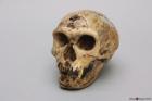
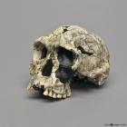
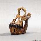
Model Nean Skull La Chapelle-Aux-Saints, 50,000 YA, Neanderthal skull was found by A. And J. Bouyssomie and J. Bonneval in 1908 in La Chapelle-aux-Saints, France. It was the most complete Neanderthal skull found at the time, Size: 22.3L x 15W x 17H (cm)
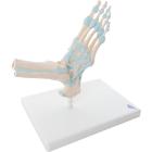
Model Foot Skeleton With Ligaments, Dimensions: 23 x 18 x 30 cm, onsists of the foot bone and lower portions of the tibia and fibula, including the introsseous membrane found between them, important ligaments and tendons of the foot are shown, large and small
Model Australopithecus afarensis Lucy Lt Finish Skull, 3.2 MYA, was discovered by D. Johanson in 1974 in Hadar, Ethiopia. Light Finish, Size: 16.6L x 13.1W x 14.5H (cm)
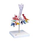
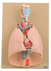
Model Ct Bronchial Tree, with Larynx and Transparent Lung, Dimensions: 22x18x37 cm, Weight: 1.905 kg, created on the basis of computer tomography data of a human, The larynx with hyoid bone and epiglottis and the trachea with primary and lobar
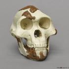
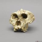
Model A. Robustus Sk-48 Cranium, 1.5-2 MYA, was discovered by Fourie in South Africa in 1950. SK-48, formerly Paranthropus crassidens, greatly increased what is known about australopithecines, Size: 15.6L x 15.1W X 12.3H (cm)
Model Homo Habilis Knm-Er 1813 Cranium, 1.9 MYA, was discovered by K. Kimeu in 1973 at Koobi Fora, Kenya, and described by R. Leakey in Nature in 1973, Size: 17.1L x 11.8W x 11.6H (cm)
Model Sivapithecus Indicus Skull, 8.5-12.5 MYA, was discovered in 1979 by Pilbeam and Shah in Pakistan. This specimen consists of a nearly complete mandible and the left side of the face, Size: 13.4L x 11.3W x 15H (cm)
Heart and lungs 7 part on plaque numbered with english keycard, Showing larynx 2-parts removable, wind pipe with bronchial tree, heart 2-parts, removable, subclavian artery and vein, venacava, aorta, pulmonary artery, oesophagus, 2 lungs front halves removable and diaphragm. On base.
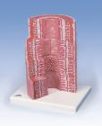
Model showing ultra structure of human heart muscle. Mounted on base. Numbered with English Key Card.Size 33 x 23 x 38 cm approx. Weight 1.8 kg approximately
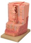
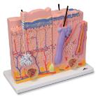
Skin 3 Part Model, The model consists of three individual parts on a common stand that represent sections of the human skin with a magnification of 80x
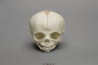
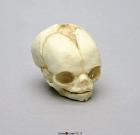
Model Fetal Human Skull 30 Weeks, Scientific Name: Homo sapiens, skull exhibits characteristics of prenatal development, 1-part skull (jaw glued to cranium), Size: 3in L x 2-1/2in W x 2-1/2in H
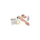
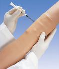
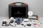
Arterial Stick Arm, Infusible arteries designed for training the proper arterial puncture procedure for blood gas analysis, For Nursing Kelly, SimPad capable
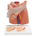
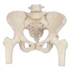
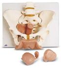
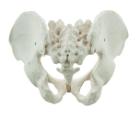
It Illustrate the morphological distinctions between male and female pelvic structures. Each pelvis includes the left and right innominates with pubic symphysis, 4th and 5th lumbar vertebrae with intervertebral discs, the sacrum and coccyx.
Model Fetal Human Skull 21 1/2 Weeks, Scientific Name: Homo sapiens, skull exhibits characteristics of prenatal development, 1-part skull (jaw glued to cranium), Size: 2-1/2in L x 1-3/4in W x 1-3/4in H