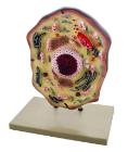
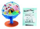
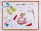
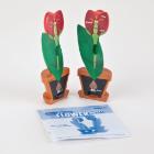
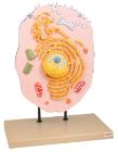
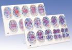
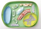
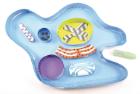
Enlarged 20,000 times. The model shows delicate structure of an animal cell. Features organelles nucleus, endoplasmic reticulum, mitochondria, ribosomes respectively polysomic and Golgi apparatus. Also showing centrioles, lysosomes and fat vacuoles.
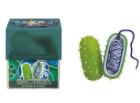
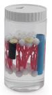
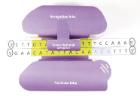
Bacteria Model, Hands-on interactive model provides a magnified and cross-sectioned detailing of Bacteria, Able to be separated, with both halves filled with a cross section of genetic material and flagellum, With Guide
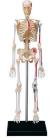
Human Anatomy Skeleton Model 4D, 9.5in model contains 46 detachable, hand-painted medical education-quality organs and parts and a display stand, Also includes illustrated assembly guide and description, For ages 8 plus
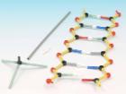
Orbit Small Dna Model Class Kit, Helps students understand DNA, Contains twelve model kits and instructions for guided learning, Students make models of 6 base pairs each that can be joined together to make a molecule of up to 72 base pairs
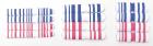
Mitosis Model, Dimensions: approx. 60x40x6 cm3, Advantages: Chromosomes coloured according to modified AZAN staining colours, Cell components are colour-coded in accordance with educational aspects, Attaching magnets on the rear, Storage system, Enlarged 10, 000 times
Three dimensional model showing electron microscopic structure. Organs like nucleus, Nucleolus, endoplasmic reticulum, mitochondria, ribosomes respectively polysomes and golgi apparatus. Showing centrioles, lysosomes and vacuoles.
Meiosis Model, Dimensions: 60 x 40 x 6 cm, Advantages: Chromosomes coloured according to modified AZAN staining colour, Cell components are colour-coded in accordance with educational aspects, Attaching magnets on the rear, Storage system, Enlarged 10, 000 times