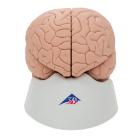
Model, Brain, 2 part, Contrasting colors are used to indicate various anatomic structures in the human brain, making this high quality model perfect for beginning anatomy studies, Made of unbreakable vinyl, Dimensions: 15 x 14 x 17.5 cm, weight: 0.82 kg
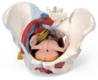
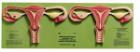
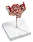
Female Pelvis with Ligaments 6 Part, This life size six part model of a female pelvis represents detailed information about the topography of bones, ligaments, vessels, nerves, pelvic floor muscles and female genital organs.
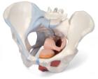
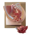
Female Pelvis with Ligaments 4 Part, This life size four part model of a female pelvis represents detailed information about the topography of bones, ligaments, pelvic floor muscles and female pelvic organs.
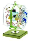
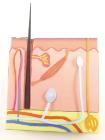
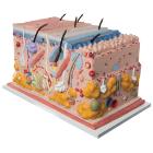
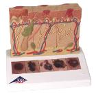
Skin 3-D Model Kit-10 Student Model Template Sets and Teacher Guide, Vinyl Pouch, Assemble and use 3-D models to visualize and investigate key science structures, Perfect for use in the classroom, lab or at home, Satisfies NGSS Standards, Grade: 6 to 10, Material: Paper, LxW: 12X19in
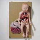
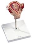
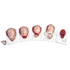
Model, Embryo, 2 month, The embryo model shows an embryo at the stage of the first month of pregnancy, Ideal for medical training and as an educational tool for pregnant women, Important anatomical structures are labeled, weight: 0.38 kg
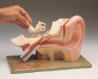
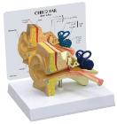
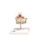
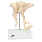
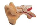
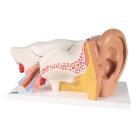
Model Ossicles Magnified 20 Times, joined to each other in the human body are located in the middle ear and are referred to as the auditory ossicles: malleus (hammer), incus (anvil) und stapes (stirrup), Weight: 0.385 kg, Dimensions : 17 x 12 x 21 cm
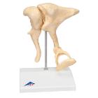
Model Ossicles 20X Bonelike, enlargement of original ossicles, created using micro CT, three smallest bones, joined to each other in the human body are located in the middle ear: malleus (hammer), incus (anvil) und stapes (stirrup), Dimensions : 17 x 12 x 21 cm
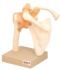
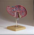
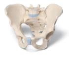
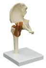
Male Pelvis, 3-Part, This 3-part model is a natural cast of a male, bone pelvis. It shows all anatomical structures in detail: both hip bones, pubic symphisis, sacrum and coccyx as well as the fifth lumbar vertebra with intervertebral disc
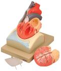
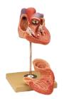
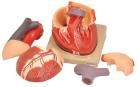
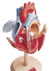
Enlarged, sectioned so that both ventricles and atria open to expose the valves. Large blood vessels near the heart and musculature of the heart are shown. Separates into 4 parts. On base.
Model, Embryo, 1 month, The embryo model shows an embryo at the stage of the first month of pregnancy, Ideal for medical training and as an educational tool for pregnant women, Important anatomical structures are labeled, weight: 0.33 kg
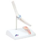
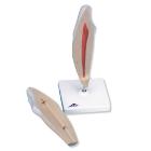
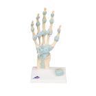
Model Hand Skeleton, with Ligaments and Carpal Tunnel, Dimensions: 30 x 14 x 10 cm, shows the anatomical detail of the ligaments and tendons found in the hand, wrist, and lower forearm, The interosseous membrane between the radius and ulna with the bones
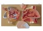
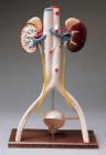
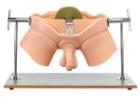
Simulator Urological Practise Phantom, Made of special plastic, allows rectal examinations and palpation of the testicles to be learned under true-to-life conditions. 5 different changes in the prostate can be felt in the anus after insertion of the fingers
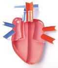
Human Heart 3-D Model Kit-10 Student Model Template Sets and Teacher Guide, Vinyl Pouch, Assemble and use 3-D models to visualize and investigate key science structures, Perfect for use in the classroom, lab or at home, Satisfies NGSS Standards, Grade: 6 to 10, Material: Paper, LxW: 12X19in