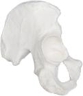
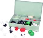
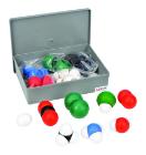
Hip Bones (Coxal Bone)(llium is part of hip bone), left, Model, Human Bone, Plastic, used as a match for your existing Eisco disarticulated skeleton, or to allow students to compare general skeletal structure
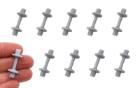
Clavicle, left (collarbone), Model, Human Bone, Plastic, These individual bones are a great addition to you collection, used as a match for your existing Eisco disarticulated skeleton, or to allow students to compare general skeletal structure
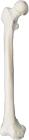
Femur, right, Model, Human Bone, Plastic, These individual bones are a great addition to you collection, used as a match for your existing Eisco disarticulated skeleton, or to allow students to compare general skeletal structure
Patella (knee cap), Model, Human Bone, Pastic, These individual bones are a great addition to you collection, used as a match for your existing Eisco disarticulated skeleton, or to allow students to compare general skeletal structure
Clavicle, right (collarbone), Model, Human Bone, Plastic, These individual bones are a great addition to you collection, used as a match for your existing Eisco disarticulated skeleton, or to allow students to compare general skeletal structure
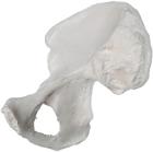
Hip Bones (Coxal Bone)(llium is part of hip bone), right, Model, Human Bone, Plastic, used as a match for your existing Eisco disarticulated skeleton, or to allow students to compare general skeletal structure
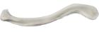
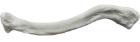
Fibula, right, Model, Human Bone, Plastic, These individual bones are a great addition to you collection, used as a match for your existing Eisco disarticulated skeleton, or to allow students to compare general skeletal structure
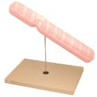
Model Ebola Virus 10000X, represents the virus magnified approximately 100000 times in its unique shape, surface shows the viral membrane with Glycoproteins, A cut-away at one end exposes internal structures major and minor matrix proteins polymerase protein and Ebola RNA
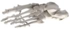
Left foot, articulated, Model, Human Bone, Plastic, These individual bones are a great addition to you collection, used as a match for your existing Eisco disarticulated skeleton, or to allow students to compare general skeletal structure
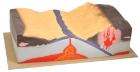
Tectonics Model, provide students with an innovative look at plate tectonics with this hand-painted plastic model, showing detailed representations of the earth layers. Both active and passive margins are shown, Oceanic floor spreading is also well-characterized
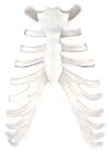
Sternum (breast bone), Model, Human Bone, Pastic, These individual bones are a great addition to you collection, used as a match for your existing Eisco disarticulated skeleton, or to allow students to compare general skeletal structure
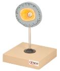
Didactic Mini Skull Model, Natural Color, Reduced size (1/2 natural size), anatomically correct replica, Realistic details, texture and bony landmarks make this model ideal for educational demonstration, Skull features natural colored details, 15 Pieces
Sacrum, Model, Human Bones, Plastic, These individual bones are a great addition to you collection, used as a match for your existing Eisco disarticulated skeleton, or to allow students to compare general skeletal structure
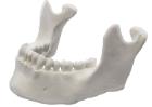
Mandible, lower jar, with 16 teeth, Model, Human Bone, Pastic, These individual bones are a great addition to you collection, used as a match for your existing Eisco disarticulated skeleton, or to allow students to compare general skeletal structure
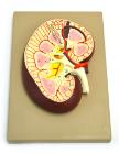
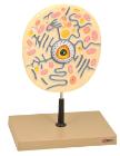
Longitudinal section of the right kidney. Model shows kidney glomerulus, tubes, one collection tube, pyramids, kidneys orifice system, kidney pelvis, upper section of the ureter and the kidney blood vessels
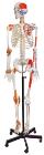
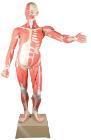
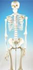
Human Skeleton Model, with Ligament Details, Painted Muscle Origins and Insertions, Flexible Spine and Joints - Rod Mount with Rolling Base, Numbered keycard, anatomical chart and dust cover included
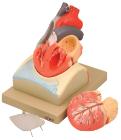
Enlarged, sectioned so that both ventricles and atria open to expose the valves. Large blood vessels near the heart and musculature of the heart are shown. Separates into 4 parts. On base.
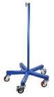
Support base for skeleton, features a heavy (30 pound) cast iron base with 5 machine grade casters (2 with locks) and a sturdy threaded pole, this skeleton stand will pass the test of time and make the transport of y skeleton easy. Inner diameter of stand is 12.5mm.
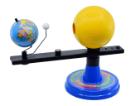
Sun, Earth, Moon Orbital Model, Multiple moving axis to demonstrate daylight, night, seasons and phases of moon, Requires two AA batteries, Light bulb inside sun to demonstrate sun light on moon & earth, Base has the months of the year along with the season
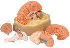
Each part on the skull is numbered. Jaw is spring loaded. Skull cap is removable for further interior inspection. Included with the model is a sheet with each numbers name for identification.
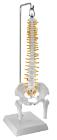
Hanging Spinal Column Model, with Pelvis & Femur Detail, Includes base and rod mount hanger, Model can be freely rotated while mounted, Model (not including mount) measures 16inch (41 cm) long, and approximately 6inch (15.25 cm) wide, Size: 1/2
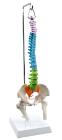
Hanging Didactic Colored Spinal Column Model, with Pelvis & Femur Detail, Includes base and rod mount hanger. Model (not including mount) measures 16inch (41 cm) long, and approximately 6inch (15 cm) wide, Size: 1/2
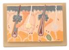
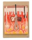
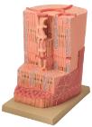
Enlarged 70 Times, full size, shows section through three layers of the hair covered skin of the head. Also shows hair follicles with sebaceous, sweat glands, receptors, nerves and vessels.
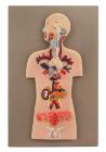
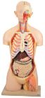
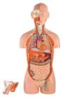
Torso model, hand painted, consists of 13 parts, removeable parts: eye with optical nerve, half brain, lung (2 parts), heart (2 parts), liver, stomach, front portion of the kidney, half of the bladder, large and small intenstines, with appendix flap opening, All parts labeled
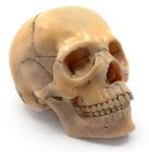
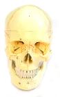
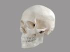
Skull Model, Human, realistic life-size, Moveable Lower Jaw, Removable Skull Cap, human skull Shows all of the bones and major structures of an adult human skull, including the fusions between bones and cartilaginous regions, Dimensions: 20L x 20W x 15H cm
Comprehensive model 300 times, enlarged vertical section showing the three layers, sebaceous and sweet glands, the hair follicles, erector muscles, arteries, nerves and veins. With key card
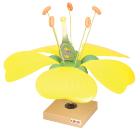
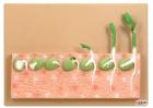
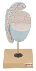
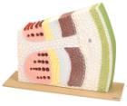
Model shows the transverse and longitudinal section of a dicotyledonous stem in which case the cambium ring has been formed but no secondary growth has taken place.
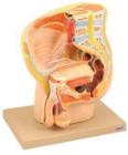
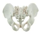
It Illustrate the morphological distinctions between male and female pelvic structures. Each pelvis includes the left and right innominates with pubic symphysis, 4th and 5th lumbar vertebrae with intervertebral discs, the sacrum and coccyx.
Model showing ultra structure of human heart muscle. Mounted on base. Numbered with English Key Card.Size 33 x 23 x 38 cm approx. Weight 1.8 kg approximately
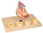
Heart dissected in two parts shows the thickened ventricle walls and cardiac valves. Four individual blood vessel model show coronary artery disease/ atherosclerosis and progression in the blood vessel, myocardial infarction and damage.