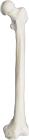
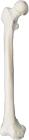
Femur, right, Model, Human Bone, Plastic, These individual bones are a great addition to you collection, used as a match for your existing Eisco disarticulated skeleton, or to allow students to compare general skeletal structure
Fibula, left, Model, Human Bone, Plastic, These individual bones are a great addition to you collection, used as a match for your existing Eisco disarticulated skeleton, or to allow students to compare general skeletal structure
Fibula, right, Model, Human Bone, Plastic, These individual bones are a great addition to you collection, used as a match for your existing Eisco disarticulated skeleton, or to allow students to compare general skeletal structure
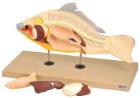
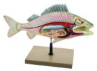
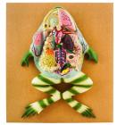
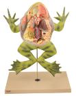
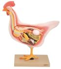
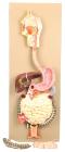
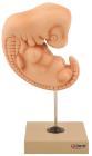
Model showing life like sculpting and vibrant hand painting faithfully portray ten organ systemsr, with numbered features. Molded of non breakable plastic base, bull frog lifts off its plastic stand for hands on study
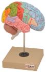
Life size, half of human brain. Students can study regions within the cerebral cortex are allied to certain functions. Impulses from the sensory organs, the skeletal muscles, skin and joints all travel to areas specialized in interpreting the information
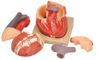
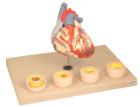
Heart dissected in two parts shows the thickened ventricle walls and cardiac valves. Four individual blood vessel model show coronary artery disease/ atherosclerosis and progression in the blood vessel, myocardial infarction and damage.
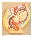
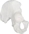
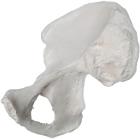
Hip Bones (Coxal Bone)(llium is part of hip bone), left, Model, Human Bone, Plastic, used as a match for your existing Eisco disarticulated skeleton, or to allow students to compare general skeletal structure
Hip Bones (Coxal Bone)(llium is part of hip bone), right, Model, Human Bone, Plastic, used as a match for your existing Eisco disarticulated skeleton, or to allow students to compare general skeletal structure
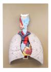
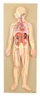
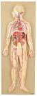
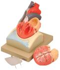
This half-size relief model helps in understand the circulation of blood in body through brain, heart, lung, liver, spleen, kidneys and partial skeleton. Mounted on base. Numbered with key card.
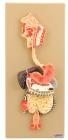
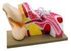
Life size model showing nose, mouth cavity and pharynx, liver with gall bladder, pancreas and spleen. Transverse colon and front stomach wall are removable. Mounted on base. Numbered with Key Card
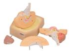
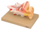
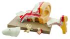
Enlarged approximately 4 times. The petros portion of the temporal bone section of the auditory canal are removable, with labyrinth can be taken out and opened. The tympanic membrane with malleus and incus can be removed in 5 parts.
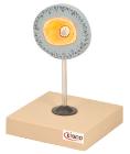
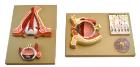
Model shows both sides of an eye, enlarged 5 x. One side of the model shows the eye socket with a sagittal cutaway and the background to the eye and the electron microscopic fine structure of the retina are shown separately.
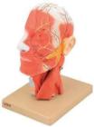
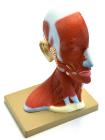
Life size model showing the outer superficial muscles, vessels, nerves and head with muscles on one side. On the outer side details of median section such as brain, mouth, larynx are shown. Includes key card.
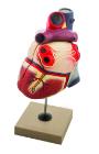
Enlarged, sectioned so that both ventricles and atria open to expose the valves. Large blood vessels near the heart and musculature of the heart are shown. Separates into 4 parts. On base.
Life size model dissectible in 2 parts. The anterior heart wall can be removed to show the left and right ventricles and atria as well as the tricuspid, pulmonary, mitral and aortic valves. Mounted on base.