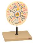
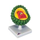
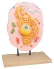
Enlarged 20,000 times. The model shows delicate structure of an animal cell. Features organelles nucleus, endoplasmic reticulum, mitochondria, ribosomes respectively polysomic and Golgi apparatus. Also showing centrioles, lysosomes and fat vacuoles.
Three dimensional model showing electron microscopic structure. Organs like nucleus, Nucleolus, endoplasmic reticulum, mitochondria, ribosomes respectively polysomes and golgi apparatus. Showing centrioles, lysosomes and vacuoles.
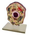
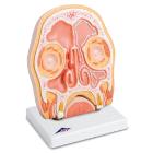
Model Head Section, Frontal section through the human paranasal sinuses covered with mucous membrane. Signs of sinusitis (paranasal sinus inflammation) on the right, with normal ventilation of the left side, Weight: 1.45 kg, Dimensions : 41 x 31 x 5 cm
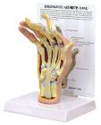
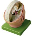
Proportions of the dura mater, natural size, made in somso-plast*, showing the proportions of the dura mater and the sinus. the 12 pairs of cranial nerves and the basilar artery with branchings are exposed. on a green base, in 2 parts. height: 23 cm, width: 18cm, depth: 21cm, weight: 900g
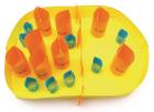
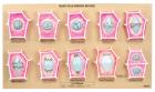
Mitosis Model, Dimensions: approx. 60x40x6 cm3, Advantages: Chromosomes coloured according to modified AZAN staining colours, Cell components are colour-coded in accordance with educational aspects, Attaching magnets on the rear, Storage system, Enlarged 10, 000 times