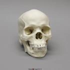
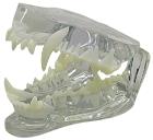
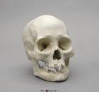
Model Human Male African-American Skull, a depressed nasal root, obtuse nasal angle, short anterior nasal spine, bilateral gutter at lower part of nasal aperture, rectangular-shaped palate & blade-like incisor in upper jaw, Size: 20.9x13.7x20cm
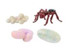
Ant Life Cycle Stages, Children can see how ants change as they grow with Insect Lore’s Ant Life Cycle Stages. These oversized, anatomically correct figures have been accurately painted and sculpted to show the four stages of ant development: eggs, larva, pupa and adult.
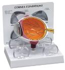
Model shows both sides of an eye, enlarged 5 x. One side of the model shows the eye socket with a sagittal cutaway and the background to the eye and the electron microscopic fine structure of the retina are shown separately.
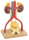
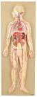
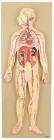
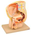
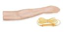
Three dimensional model of male urinary system and its blood supply is free standing, enabling study from all aspects. The kidneys, adrenal & blood vessels, a section of the pelvic bone with pubic symphysis, lower portion of the bladder and prostate
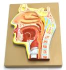
Enlarged 2 times, this cross section of human head to show mouth and nasal passages. Relationship between the windpipe and esophagus can be easily shown. Includes key card
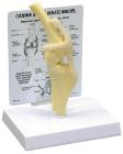
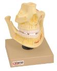
This model of adult upper and lower jaw shows tooth roots, spongiosa, vessels and nerves are exposed. The lower jaw is movable with the help of spring action.
Enlarged approximately 4 times. The petros portion of the temporal bone section of the auditory canal are removable, with labyrinth can be taken out and opened. The tympanic membrane with malleus and incus can be removed in 5 parts.
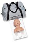
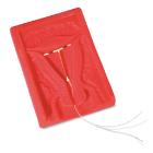
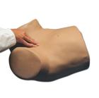
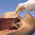
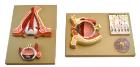
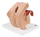
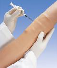
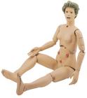
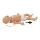
Model For Gynecological Patient Education, This unique gynecological training model is ideal for demonstration purposes and for realistic insertion of female barrier contraceptive devices, which are placed in the vaginal/cervical area.
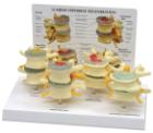
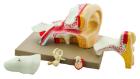
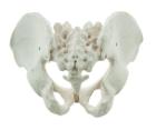
It Illustrate the morphological distinctions between male and female pelvic structures. Each pelvis includes the left and right innominates with pubic symphysis, 4th and 5th lumbar vertebrae with intervertebral discs, the sacrum and coccyx.
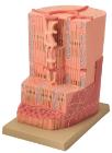
Model showing ultra structure of human heart muscle. Mounted on base. Numbered with English Key Card.Size 33 x 23 x 38 cm approx. Weight 1.8 kg approximately
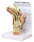
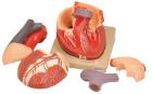
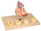
Heart dissected in two parts shows the thickened ventricle walls and cardiac valves. Four individual blood vessel model show coronary artery disease/ atherosclerosis and progression in the blood vessel, myocardial infarction and damage.
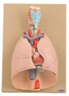
Heart and lungs 7 part on plaque numbered with english keycard, Showing larynx 2-parts removable, wind pipe with bronchial tree, heart 2-parts, removable, subclavian artery and vein, venacava, aorta, pulmonary artery, oesophagus, 2 lungs front halves removable and diaphragm. On base.
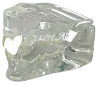
Model Human Female American Indian Skull, Includes broad, flattened, forward-projecting zygomas, facial bones are vertically aligned with shallow nasal depression, moderate nasal spine, orthognathic jaw & vertical chin, Size: 18.5x14x18.8cm
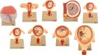
Set of nine models, showing the following stages. 1. Embryo 6 days old 2. 1st month of gestation. 3. Uterus with embryo in 3rd month of gestation.4. Uterus with fetus, in 4th month. 5. Uterus with fetus, placenta and umbilical cord.6. 5th month. 7. 7th month pregnancy.