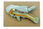
Amoeba proteus, enlarged approximately 1000 times. In a small pseudopodium which can be opened up showing the structure after electron microscopic magnification. On a base with explanatory notes. Separates into 2 parts
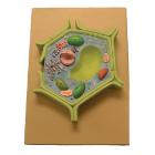
This plant cell model is separated in 4 parts. Showing details of cell wall and inner details of cell wall, nucleus is separated and defined in a 3-D way cut section of chloroplast is shown.
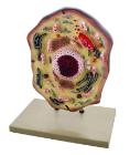
Enlarged 20,000 times. The model shows delicate structure of an animal cell. Features organelles nucleus, endoplasmic reticulum, mitochondria, ribosomes respectively polysomic and Golgi apparatus. Also showing centrioles, lysosomes and fat vacuoles.
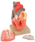
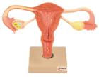
Sectioned through the ventricles and auricles. The bicuspid and tricuspid semilunar and sigmoid valves are shown. Separates into 3 parts. Mounted on base
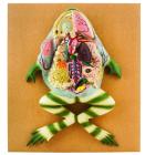
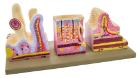
Life size model showing nose, mouth cavity and pharynx, liver with gall bladder, pancreas and spleen. Transverse colon and front stomach wall are removable. Mounted on base. Numbered with Key Card
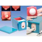
Birthing Stages Trainer, contents: 6 birth stage inserts, Safety plate with 4 screws (for the optional insert in birth simulator P90), Lubricant, Carry bag, The inserts look the same from the outside, using a number code on the back, the condition of the cervix on the inside
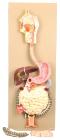
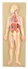
This half-size relief model helps in understand the circulation of blood in body through brain, heart, lung, liver, spleen, kidneys and partial skeleton. Mounted on base. Numbered with key card.
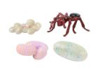
Ant Life Cycle Stages, Children can see how ants change as they grow with Insect Lore’s Ant Life Cycle Stages. These oversized, anatomically correct figures have been accurately painted and sculpted to show the four stages of ant development: eggs, larva, pupa and adult.