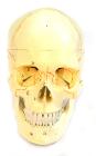
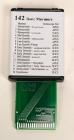
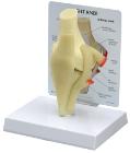
Each part on the skull is numbered. Jaw is spring loaded. Skull cap is removable for further interior inspection. Included with the model is a sheet with each numbers name for identification.
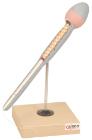
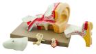
Enlarged approximately 4 times. The petros portion of the temporal bone section of the auditory canal are removable, with labyrinth can be taken out and opened. The tympanic membrane with malleus and incus can be removed in 5 parts.
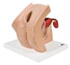
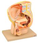
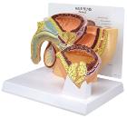
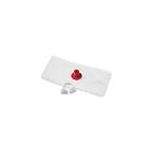
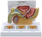
Model For Gynecological Patient Education, This unique gynecological training model is ideal for demonstration purposes and for realistic insertion of female barrier contraceptive devices, which are placed in the vaginal/cervical area.
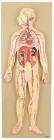
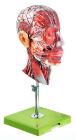
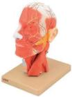
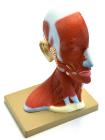
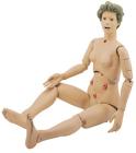
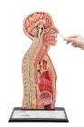
Life size model showing the outer superficial muscles, vessels, nerves and head with muscles on one side. On the outer side details of median section such as brain, mouth, larynx are shown. Includes key card.
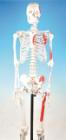
Life size skeleton features the points of origin in red and the points of insertion in blue along the left side. Skeleton features a sectioned calvarium and hinged jaw. Includes rod mounted stand with casters. Hands form ring on top of skull
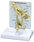
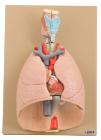
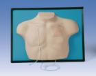
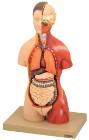
Heart and lungs 7 part on plaque numbered with english keycard, Showing larynx 2-parts removable, wind pipe with bronchial tree, heart 2-parts, removable, subclavian artery and vein, venacava, aorta, pulmonary artery, oesophagus, 2 lungs front halves removable and diaphragm. On base.
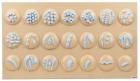
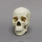
Model Human Female Asian Skull, include a flat nasal root, an obtuse nasal angle, a somewhat rounded dental arcade in the upper jaw, mild to moderate prognathism and shovel-like incisors in the upper jaw, Size: 19.4L x 12.9W X 17.3H (cm)
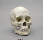
Model Human Female American Indian Skull, Includes broad, flattened, forward-projecting zygomas, facial bones are vertically aligned with shallow nasal depression, moderate nasal spine, orthognathic jaw & vertical chin, Size: 18.5x14x18.8cm
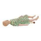
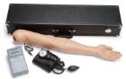
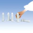
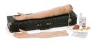
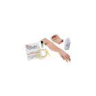
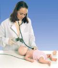
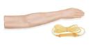
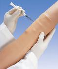
Arterial Stick Arm, Infusible arteries designed for training the proper arterial puncture procedure for blood gas analysis, For Nursing Kelly, SimPad capable
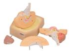
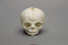
Model Fetal Human Skull 30 Weeks, Scientific Name: Homo sapiens, skull exhibits characteristics of prenatal development, 1-part skull (jaw glued to cranium), Size: 3in L x 2-1/2in W x 2-1/2in H
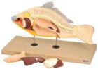
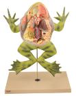
Model showing life like sculpting and vibrant hand painting faithfully portray ten organ systemsr, with numbered features. Molded of non breakable plastic base, bull frog lifts off its plastic stand for hands on study
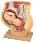
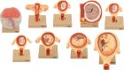
Set of nine models, showing the following stages. 1. Embryo 6 days old 2. 1st month of gestation. 3. Uterus with embryo in 3rd month of gestation.4. Uterus with fetus, in 4th month. 5. Uterus with fetus, placenta and umbilical cord.6. 5th month. 7. 7th month pregnancy.
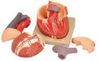
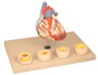
Heart dissected in two parts shows the thickened ventricle walls and cardiac valves. Four individual blood vessel model show coronary artery disease/ atherosclerosis and progression in the blood vessel, myocardial infarction and damage.
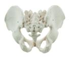
It Illustrate the morphological distinctions between male and female pelvic structures. Each pelvis includes the left and right innominates with pubic symphysis, 4th and 5th lumbar vertebrae with intervertebral discs, the sacrum and coccyx.
Model showing ultra structure of human heart muscle. Mounted on base. Numbered with English Key Card.Size 33 x 23 x 38 cm approx. Weight 1.8 kg approximately
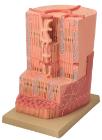
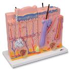
Skin 3 Part Model, The model consists of three individual parts on a common stand that represent sections of the human skin with a magnification of 80x
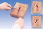
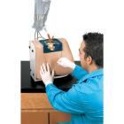
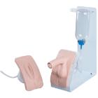
BASIC catheterization trainer set, both male and female bladder catheterization procedures can be realistically demonstrated, practiced and assessed. The easy-to-change genital inserted into the holder and held in place with magnets, with Both inserts
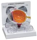
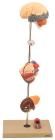
Showing the harmful effects of hypertension on the most susceptible organs. It consists of scaled down depictions of brain, eye, 2-part heart, 2-part kidney, enlarged artery. All of the organs can be rotated or removed for closer viewing.
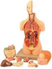
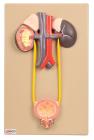
Natural size. Kidneys, ureters, adrenal glands and bladder with prostate as well as the large abdominal right kidney sectioned to show all anatomical details.
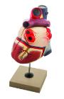
Life size model dissectible in 2 parts. The anterior heart wall can be removed to show the left and right ventricles and atria as well as the tricuspid, pulmonary, mitral and aortic valves. Mounted on base.