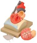
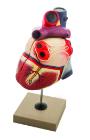
Enlarged, sectioned so that both ventricles and atria open to expose the valves. Large blood vessels near the heart and musculature of the heart are shown. Separates into 4 parts. On base.
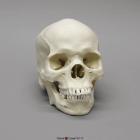
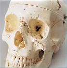
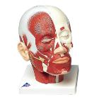
Model Human Male African-American Skull, a depressed nasal root, obtuse nasal angle, short anterior nasal spine, bilateral gutter at lower part of nasal aperture, rectangular-shaped palate & blade-like incisor in upper jaw, Size: 20.9x13.7x20cm
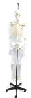
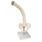
Support base for skeleton, features a heavy (30 pound) cast iron base with 5 machine grade casters (2 with locks) and a sturdy threaded pole, this skeleton stand will pass the test of time and make the transport of y skeleton easy. Inner diameter of stand is 12.5mm.
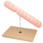
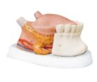
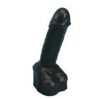
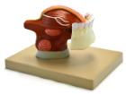
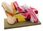
Enlarged 3x, shows the male urinary bladder with the prostate gland surrounding the urethra. The model is dissected medially to expose both inteRNAl and exteRNAl structures of the bladder and prostate
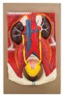
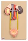
Model Human Urinary Organs, Weight (lb):2.6, Length (in):13.0,Width (in): 9.1, Height (in): 4.3,Natural Size separates into 3 parts. Kidneys ureters, adrenal glands, bladder with prostate and major blood vessels
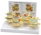
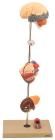
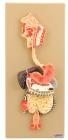
Showing the harmful effects of hypertension on the most susceptible organs. It consists of scaled down depictions of brain, eye, 2-part heart, 2-part kidney, enlarged artery. All of the organs can be rotated or removed for closer viewing.
Natural size. Kidneys, ureters, adrenal glands and bladder with prostate as well as the large abdominal right kidney sectioned to show all anatomical details.
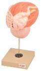
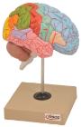
Life size, half of human brain. Students can study regions within the cerebral cortex are allied to certain functions. Impulses from the sensory organs, the skeletal muscles, skin and joints all travel to areas specialized in interpreting the information
Life size model dissectible in 2 parts. The anterior heart wall can be removed to show the left and right ventricles and atria as well as the tricuspid, pulmonary, mitral and aortic valves. Mounted on base.
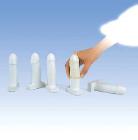
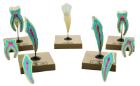
Lower incisor with removable half of crown, lower canine separates in two longitudinally, lower molar with one root is one-piece and two roots separate into three parts, upper molar with three roots separates into three parts.
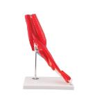
Model Elbow Joint 8 Parts, great tool for student and patient education, part of a high quality series of muscle models, and has been manufactured to replicate the anatomy of the human elbow joint in detail, colors used, Weight: 1.74 kg, Dimensions : 25 x 41 x 25 cm
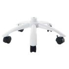
Metal base, with 5 casters, Dimensions: 53.3 x 53.3 x 17.8 cm, features extraordinary stability and durability. The 5 large casters ensure that the stand is stable and easy to handle, The base is compatible with all 3B skeletons and the 3B muscle figures B50 and B51