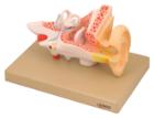
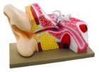
Enlarged approximately 4 times. The petros portion of the temporal bone section of the auditory canal are removable, with labyrinth can be taken out and opened. The tympanic membrane with malleus and incus can be removed in 5 parts.
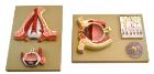
Model shows both sides of an eye, enlarged 5 x. One side of the model shows the eye socket with a sagittal cutaway and the background to the eye and the electron microscopic fine structure of the retina are shown separately.
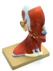
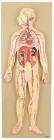
Life size model showing the outer superficial muscles, vessels, nerves and head with muscles on one side. On the outer side details of median section such as brain, mouth, larynx are shown. Includes key card.
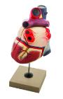
Enlarged, sectioned so that both ventricles and atria open to expose the valves. Large blood vessels near the heart and musculature of the heart are shown. Separates into 4 parts. On base.
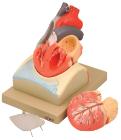
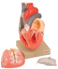
Life size model dissectible in 2 parts. The anterior heart wall can be removed to show the left and right ventricles and atria as well as the tricuspid, pulmonary, mitral and aortic valves. Mounted on base.
Sectioned through the ventricles and auricles. The bicuspid and tricuspid semilunar and sigmoid valves are shown. Separates into 3 parts. Mounted on base
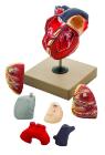
Enlarged. Sectioned so that both ventricles and atria open to expose the valves. Large blood vessels near the heart and musculature of the heart are shown. Separates into 7 parts.
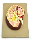
Longitudinal section of the right kidney. Model shows kidney glomerulus, tubes, one collection tube, pyramids, kidneys orifice system, kidney pelvis, upper section of the ureter and the kidney blood vessels