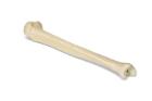
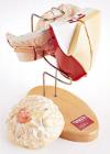
ORTHObones* Premium Model, Tibia Left, is used by surgeons, orthopaedists and medical engineers worldwide, high quality, biomechanical bones perfectly resemble a human bone, with the two distinct layers, Dimensions: 39 x 81 x 7 cm, weight: 0.216 kg
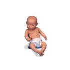
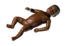
Simulator Nursing Baby, Female 6 week old baby, in SOMSO-Plast*, Black color, With ball joints, head moves easily and tilts backwards. Painted eyes, Nose and ears are open as is anus for insertion of thermometer, Undressed, for nursing exercises, Size of head 35.8 cm
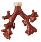
Simulator Florescing Tracheo-Bronchial Tree, for use with the Light Imaging Fluorescence Endoscope (LIFE Æ Xillix/Olympus), or SAFE (1000 Pentax), bronchial mucosa appears normal with conventional bronchoscopy Teaching Model
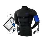
Force Transducer, high sensitivity, dual-range, research grade, designed to measure forces in the 0.005 to 10 gram and 0 to 100 gram ranges, exhibits excellent isometric properties, Sensitivity: lessthan60 mV/g (0-10 g), Noise: 3.0 mg ma
Simulator, Placenta, folios, for PPH Trainer P97 (set of 10), with 2 possible detachable pieces for the PPH Trainer P97 (1021568) and PPH Trainer P97- Module (1021567)
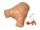
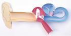
Simulator Catheterizing-Model, with interchangeable male and female genital organs as well as interchangeable bladder, in natural size, made of special plastic, Weight: 6 kg, Length: 46 cm, Width: 43 cm, Height: 23 cm
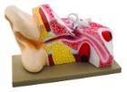
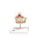
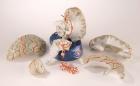
Human Ear 3-D Model Kit-10 Student Model Template Sets and Teacher Guide, Vinyl Pouch, Assemble and use 3-D models to visualize and investigate key science structures, Perfect for use in the classroom, lab or at home, Satisfies NGSS Standards, Grade: 6 to 10, Material: Paper, LxW: 12X19in
simulator, Complete abdominal wall set, for the birth simulator, P90 Pro, includes the abdominal wall and the C-section insert that can be sectioned, Dimesnions: 47 x 35 x 28 cm, Weight: 1.05 kg